
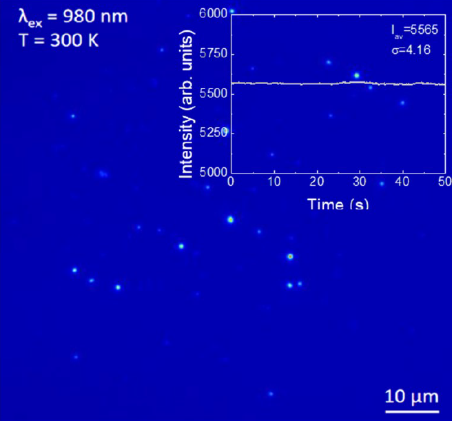
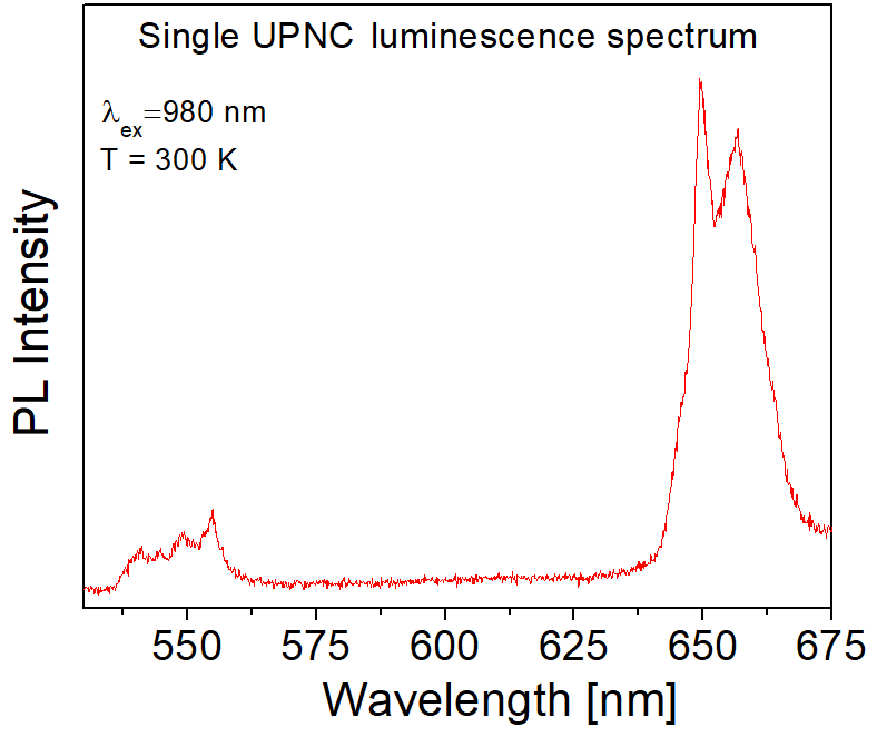
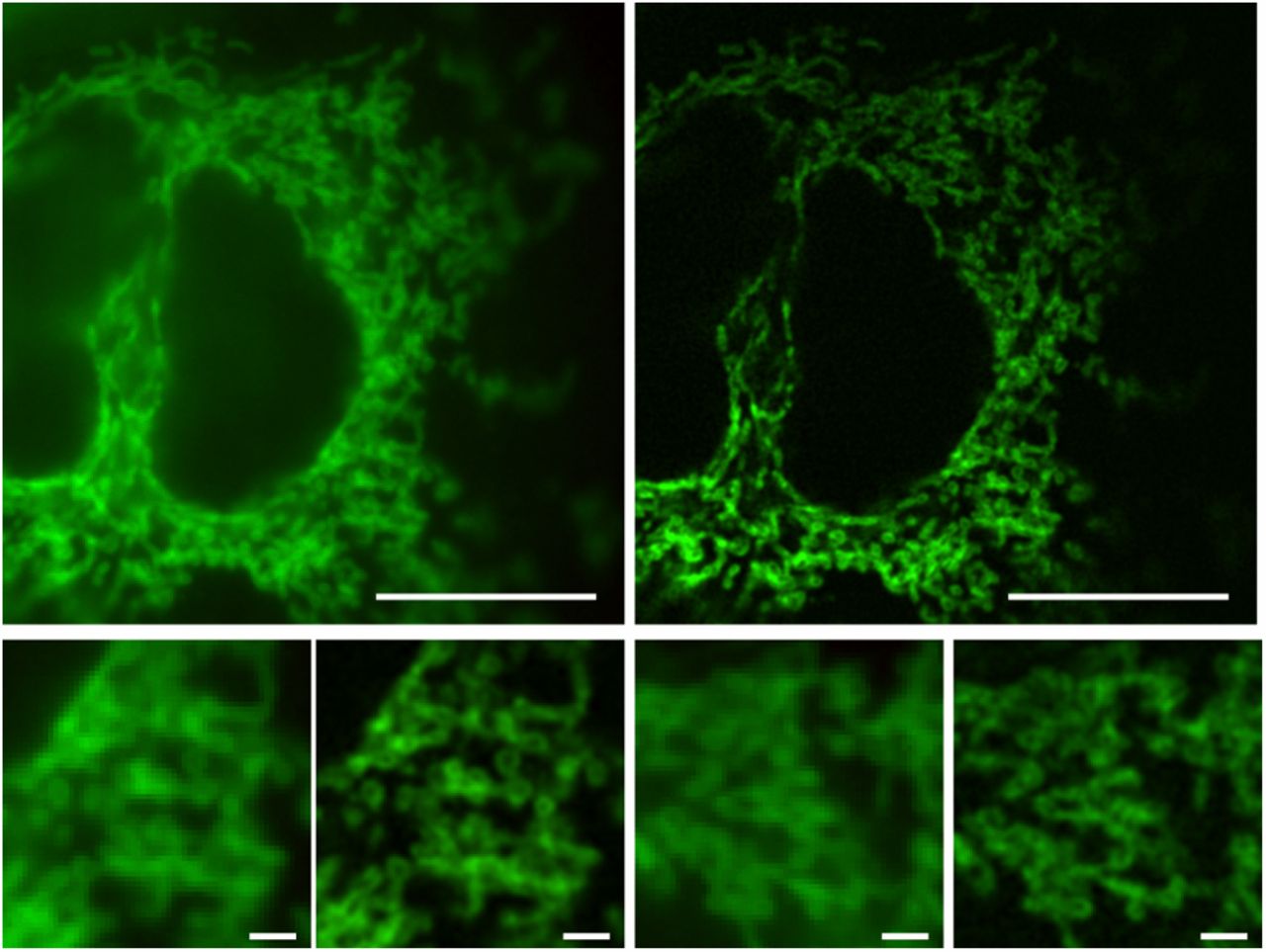
Group of Colloidal Nanostructures utilizes single fluorescence microscopy techniques to probe the optical properties of inorganic nanostructures, one nanoparticle at a time. Our main microscope is equipped with sensitive spectrometer operating in both VIS and NIR spectral region, which allows us to measure emission spectra of single nanoparticles or molecules. Due to high temporal and spatial resolution of our detectors, single quantum dots fluorescence intermittencies could be studied as well as single molecule Forster Resonance Energy Transfer (smFRET). In the case of bioimaging, we are using Super-resolution optical fluctuation imaging (SOFI) technique to circumvent the diffraction limit. SOFI algorithms are capable of image reconstruction of densely labeled samples with the resolution of tens of nanometers as well as background reduction and 3D sectioning. These approaches are applied to study the energy transfer in single upconverting nanoparticles, blinking behavior of quantum dots, energy transfer in hybrid nanostructures, metalloprotein conformational changes, bacteria dividing process.
[Dedecker et al., PNAS 2012]